 |
Non l'ho ancora iniziata la traduzione ... Scusate.
|
 |
Neonatal Hydronephrosis The
kidneys, located in the back of the abdomen just below the ribs, are important for many
bodily functions. One of the kidneys' major functions is to filter blood and remove waste
products that are passed out of the body through the urine. The kidneys are connected to
thin tubes called ureters, which carry the urine to the bladder. When the kidneys are
forming before a child is born, sometimes conditions develop that change the way one or
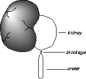
both of the kidneys look or function. One such condition is hydronephrosis. Hydronephrosis is a "stretching" or dilation of the inside or collecting part of the kidney. It often results from a blockage in the ureter where it joins the kidney that prevents urine from draining into the bladder. Urine is trapped in the kidney and causes the kidney to stretch. Hydronephrosis may also be due to abnormal backwash or "reflux" of urine from the bladder. In infants the amount of hydronephrosis may appear greater than the actual degree of blockage because of the "stretchiness" of the young tissues. Neonatal hydronephrosis is often detected on an ultrasound test during pregnancy (prenatal ultrasound). Hydronephrosis has never been linked to anything the parents have done during pregnancy and is usually not passed from parent to child or found in more than one sibling in a family. The blockage that produces hydronephrosis is usually the result of a narrowing at the top of the ureter near the kidney that probably developed before the fourth month of pregnancy. The severity of hydronephrosis depends on the extent of the blockage and the amount of
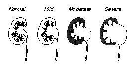
stretching of the kidney. Hydronephrosis may range from mild to severe. Children with mild
hydronephrosis usually do not have symptoms, the kidneys are minimally affected, and the
hydronephrosis may disappear in the first year of life. In patients observed with moderate
hydronephrosis, kidney function usually does not decrease, kidney growth remains normal,
and the degree of hydronephrosis usually does not worsen. Research has shown that careful
observation during the first one to two years of life suggests that the kidney compensates
for the hydronephrosis to maintain normal function and that some children even get better
on their own. In extremely severe cases of hydronephrosis, damage to normal kidney function may occur. In addition to affecting the child's kidney function, this condition may also cause infections, pain, and bleeding. These effects may take months or even years to occur or may never occur. To determine the amount of hydronephrosis present, nuclear medicine and radiologic tests to measure kidney function and structure may be recommended by the physician. In children with mild hydronephrosis, observational therapy has been shown to be safe and has become the accepted method of treatment. However, in children with moderate to severe hydronephrosis, the answer is not as clear. The surgery to correct hydronephrosis is called pyeloplasty. This procedure involves removing the obstructed part of the ureter and then reattaching the healthy ureter to the collecting part of the kidney. After the operation the child remains in the hospital, usually for 3 to 5 days. The success rate of the surgical therapy in infants is 90-95%. Yet, some studies suggest that observation is better than surgery in children with moderate to severe hydronephrosis since the problem may resolve itself without the risks of anesthesia and surgery, kidney function is not lost, and the kidneys grow well. Since 1985, prenatal testing for hydronephrosis has permitted early detection and treatment of hydronephrosis. In the past most children were found to have hydronephrosis at the time of urinary infection or pain. Surgery was almost always performed; in many children this was after 3-4 years of age. Those children who were born with hydronephrosis that improved by itself without ever causing infection or pain, were probably never diagnosed or treated for hydronephrosis. Children who are diagnosed prenatally with moderate to severe hydronephrosis are now being seen at such an early age that they have not had a chance to improve on their own. Current testing cannot accurately predict which patients might or might not get better on their own. Therefore, today there is no standard treatment for all children. Many centers are choosing to watch and carefully monitor children with moderate to severe hydronephrosis while others continue to use surgery as treatment.
|
||

